


Visit also the main downy mildew information page, DM on plants, & how to spot downy mildew on leaves
Here we look at the effect on individual plants, with photos over time. It was this spring (2014) when I first noticed some unusual new growth on a couple of my aquilegias. Being very light in colour with more 'upright-seeming' growth and long leaf stems, I wondered if it was a new & interesting variation I could start selecting for. !!! Little did I know then that it was a new, virulent disease and that it would threaten my whole aquilegia collection.
As the leaves developed, I did have a feeling that the growth looked 'wrong': the lightness and lankiness of the growth made it look etiolated, or possibly chlorotic and struggling. Growth then became more 'frazzled' looking as the leaves curled upwards or downwards, giving more of an appearance that they had distorted growth like that seen on plants affected by weedkiller. But no weedkiller had been used
T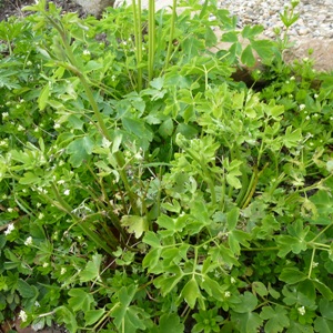 hen
both my neighbour and myself noticed the same effect on an aquilegia by
her front path. An unusual pattern of growth, lighter in colour than
even the lighter green of white-flowered aquilegias; upright and
curled/frazzled growth. (Hard to see in the photo as there is ground
cover of sweet woodruff, and another (white flowered) aquilegia directly
behind it.) I decided to take a series of photos to see how it
developed. Then results came through from
the RHS Members' Advisory Service to give me the bad news: it's downy
mildew, a new disease of aquilegias. These are signs of
systemic infection. My neighbour and I decided that I'd
continue to take photos to see what happened over time. No, I didn't set
the camera up exactly the same way each time, and, anyway, the
increasingly tall top growth meant that I had to change the viewpoint
and relative size of plant; nor am I a professional photographer, not
even using a tripod. However, I hope that this visual experiment over
time yields useful information for not only myself and my neighbour, but
also other gardeners worldwide.
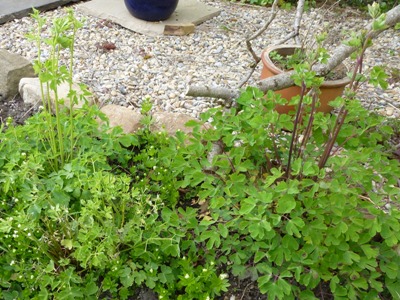
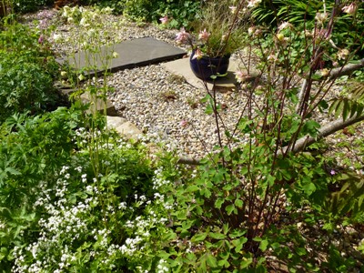
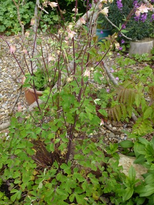
In an arbitrary but convenient way, I will designate 'Day 0' as the day the first photo was taken. Subsequent photos are then designated as 'Day X' where X is the number of days since the taking of the first photo. The affected plant was one of a group of three. Closest was a white-flowered one ....the one behind in the photo above. The other plant was further away to the right, and is pink-flowered. I'll let the photos do most of the rest of the talking:
Case study: a group of 3 plants |
||
| Here's the group of 3 plants, plus, far right, a close-up of
the systemically infected one. This is Day 0 Flowering stems are starting to shoot up, they grow very rapidly. |
 |
 |
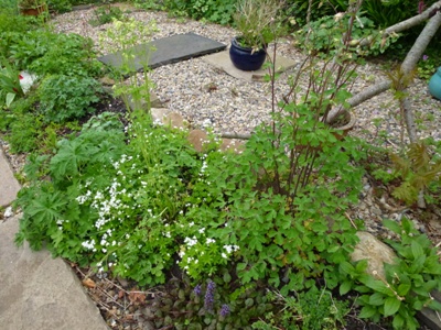
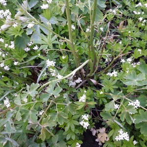
| Day 11 The almost-horizontal flower-stem is due to slug damage leading to collapse. Please graciously ignore the colour-cast seen here and on some other photos, it's an aberration of the changing camera conditions. |
 |
 |
|
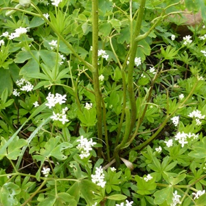
Day 15 Where are the leaves going? Many now reduced to leaf-stems with no lamina. The sweet woodruff is rapidly growing taller and denser, complicating the visual effects. The horizontal, eaten stem has broken free totally and is no longer in the photo. |
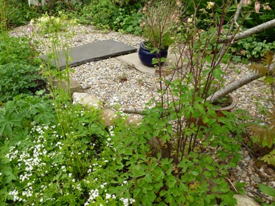 |
 |
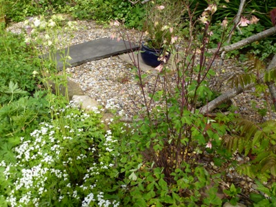
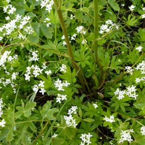
| Day 19 |
 |
 |
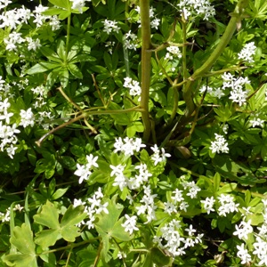
| Day 23 |
 |
 |
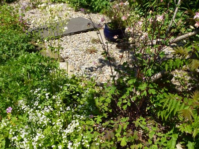
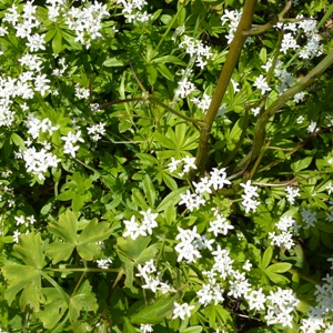
| Day 27 |
 |
 |
|
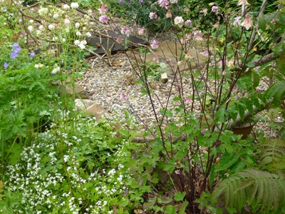
Day 31 Apart from that persistent leaf, bottom left, all that's left is leaf and flower stems, and they are dying back and becoming blackened and wiry. |
 |
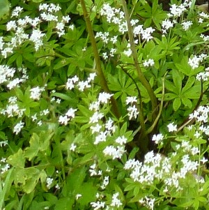 |
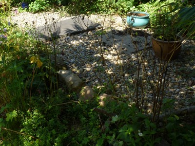
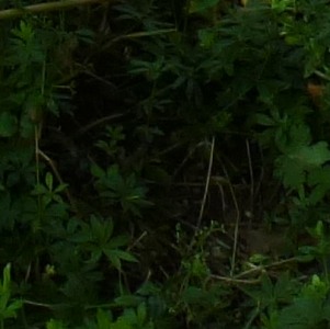
| Day 97 3 months later than the first photo, remaining plants are now setting seeds. Darn sun wouldn't stop shining! All that's left of the infected plant is a bare circle in the woodruff. The top of the roots (at soil surface) were showing for quite a long time after all the top growth had disappeared. |
 |
 |
Portrait 1: Death of an infected plant |
||
| Let's look closer at the signs in the plant that was
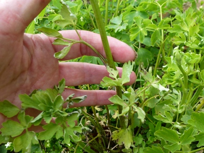
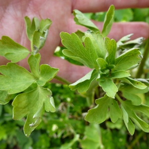
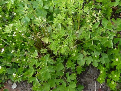
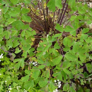
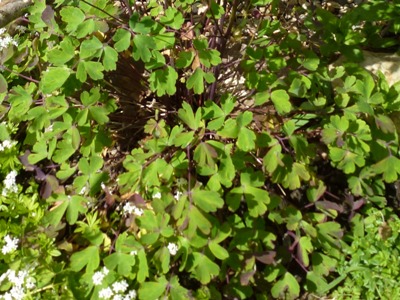
affected. Remember, this plant was systemically infected as it came into growth in the spring. Day 0 This is what I currently believe to be meristem infection: the disease has entered the growing tissue where leaves have formed. Less likely, it could be that there are spores around the growing tip that infect the new leaf tissue soon after formation, anyway, the whole leaf is affected. |
 |
 |
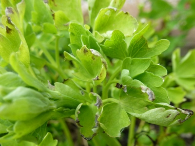
| Day 0 The leaf stems are quite long
compared to the leaf surface (lamina), and seem to grow quite
upright rather than splayed out (which ordinary aquilegia leaves
will do, in order to maximise the amount of light a leaf
receives). These look 'frazzled' because the leaf lamina is curled upwards. Furthermore, the edges of the leaves have started to look 'singed' as dead tissue results, probably, from the leaf dying back in response to the infection. Far right: more and more leaf dies away. |
 |
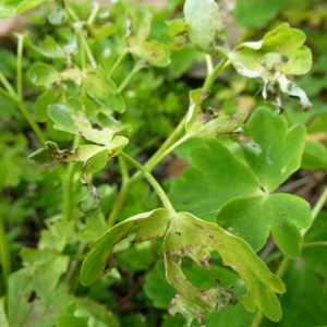 |
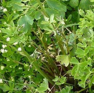
| Already, at Day 0, some shoots/noses have begun to have
disintegration of the leaves and die-back ( close-up far right). Looking closely at the main photo, we see that not all leaves are infected, especially bottom left, we see normally coloured and shaped leaves that splay out near the ground. If you revisit the right-hand photos in the table above you will see how this leaf remains in 'good' health versus the other disintegrating ones. |
 |
 |
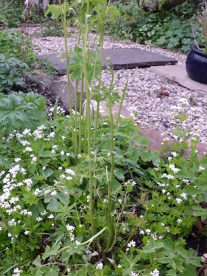
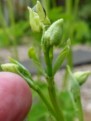
| Day 11 NB The other aquilegia behind makes it difficult to
discern which stems are which plant. Those buds look normal. However, see the little leaves under the buds? They are curling. And there is slug slime and deposit: a slug would not normally be venturing here. 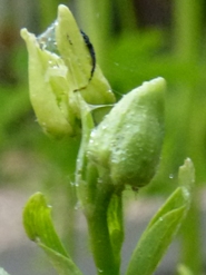 |
 |
 |
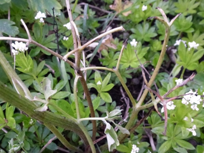
| At Day 11 many leaves have lost their surface, and the others look decidedly sick, being very curled and with much dead tissue. And spores are probably pouring from the undersides as well as being releasing by dying tissues. |
 |
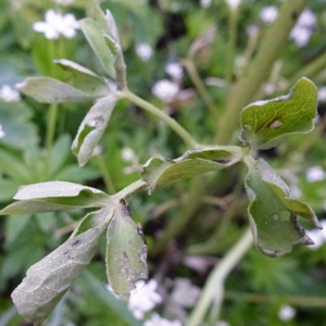 |
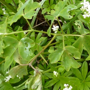
| Now at Day 15 we notice that the right hand leaf shown 3
rows above is showing signs of Downy mildew infection, note this
is different from the infection we saw in the new leaves, and
has probably resulted from infection by spores released from the
other leaves. So, far right, we see slightly yellowy patches, dying leaflets, and slug slime. There are also holes, probably caused by slug grazing in this case rather than leaf tissue death. |
 |
 |
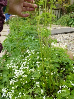
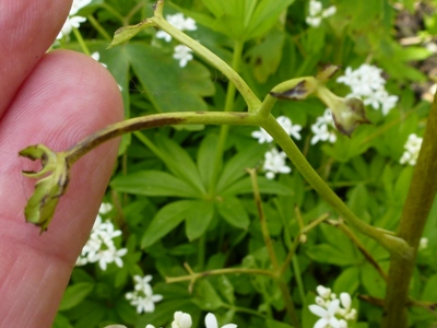
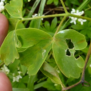
| Just 4 days later at Day 19, the camera captures how some of
the originally infected leaves have now died back so there is
almost no leaf lamina left. The leaf vein near my fingers shows
signs of infection as a brown streak. Far right, a close-up of the leaf above to show the yellow patches which are beginning to be brown-tinged, and also (probably) slug-grazed holes. |
 |
 |
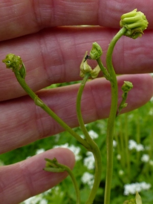
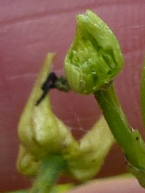
| Day 19 shows the kinky, distorted, flower stems, and infected flower buds that have been eaten by slugs or snails. |
 |
 |
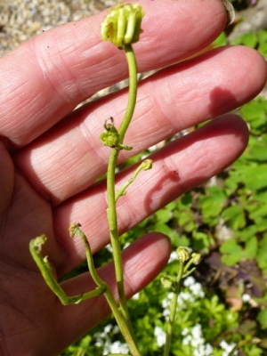
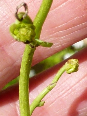
| Day 23 Bud-loss to gastropods continues. |
 |
 |
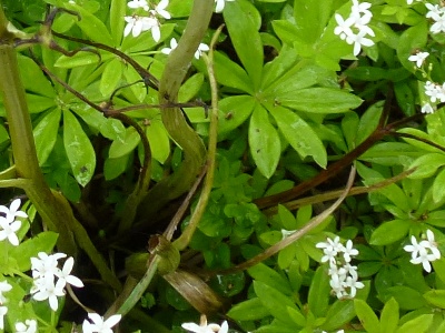
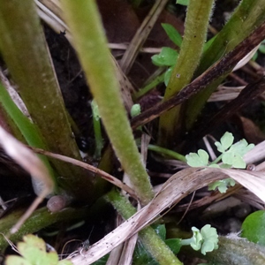
| Day 31 Infected leaves are now just spindly dead petioles. Flower stems still have some green tissue left, but never were able to produce any flowers. A few new, systemically infected, leaves are still being produced. But basically, this plant is dead. |
 |
 |
| Knowing, now, that plants infected in their new growth are likely to die (and infect other plants before doing so) I now suggest digging out such plants at the earliest opportunity. See also the page about plants whose leaves are affected in this way that I believe is meristemic infection. | ||
Portrait 2: Disease progress in a plant |
||
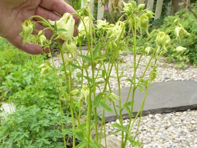
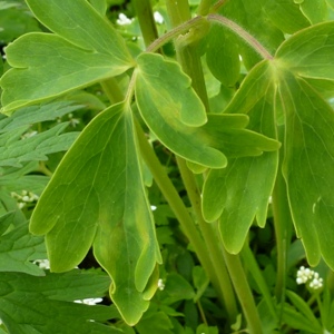
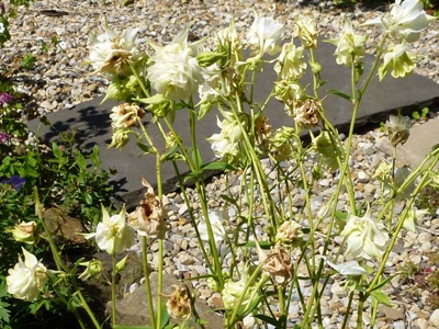
| Let's look at the white flowered aquilegia plant that was
right behind the infected one. At Day 11 the flower buds are looking good, but some leaves now start to show the signs of infection. These infected yellowy patches on the leaflets are likely from spores released by the nearby infected plant. |
 |
 |
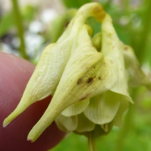
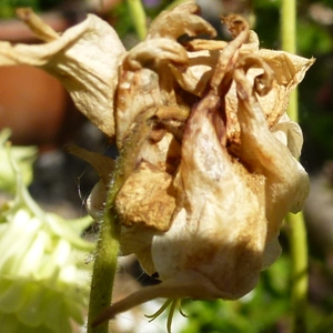
| However four days later, at Day 19, on some flowers there's the distinctive
brown dots and streaks that I believe denote downy mildew. An aphid has also set up home, and given birth to live young several times recently. |
 |
 |
| Day 27 and more flowers show infection. |
 |
 |
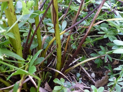
| Day 97 / 3 months. Flowering well over, this plant actually set seeds in the unaffected flowers that it produced. The remaining flower stems are showing elongated darkened areas. There is usually a browning off and dying of these stems (and leaves) at this time, but I think that these particular patches are downy mildew infection. |
 |
|
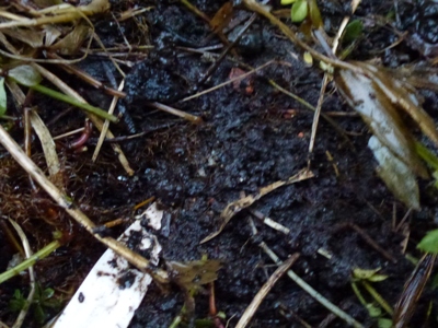
| It's now wintertime, nearly the solstice here in South
Wales, UK. Mild and wet. I went looking for the plant to get the latest photo of what is happening. I found the plant label. It looks as if the plant has died. Although sometimes a dormant aquilegia may not be very visible at all, usually they like to poke their noses up out of the soil. |
 |
|
Portrait 3: Disease progress in a plant |
||
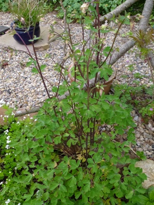
| Here's the third plant shown in the first table above. It is
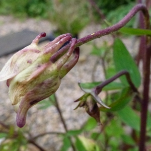
the one to the right of the group of three plants. Day 11. Nice strong plant with plenty of healthy foliage. Or is it? Closer inspection shows possibly infected leaflets on a flower-stem leaf, and browning of another leaf. Some buds are discoloured and are carried on particularly kinky stems. As there's no obvious mollusc damage to the stem (and the plant never wilted), I postulate that it could be that infected tissue shows abnormal response to gravity, so causing the strange distortion. |
 |
 |
At 15 days, I spot an unusual white-silver effect on a leaf.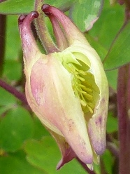 |
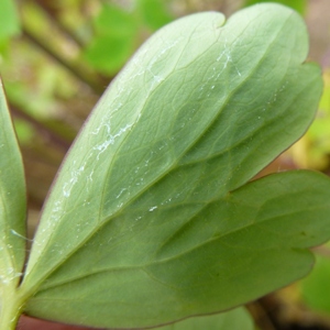
There are also browner areas though I cannot find any
of the typical yellowy areas on any leaf. The underside of the
leaflet shows no fungal growth, although slugs have shown an
interest, and also have eaten part of a bud.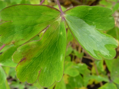 |
 |
By 19 days this plant is still looking almost unaffected
except for the flowers and upper flowering stems. The leaves are
strong and healthy and most of the dark
discoloration seen is unlikely to be DM.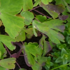 |
 |
 |
| Day 23 |
 |
|
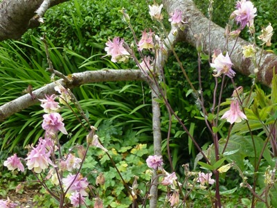
| At Day 31 there's still good flowering with most flowers uninfected, but the infected ones are very browned and dried looking. |
 |
|
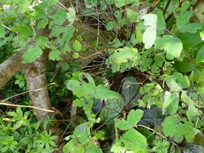
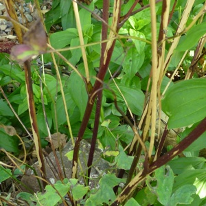
| Day 97 (3months) Leaves look very bedraggled and holey, although it doesn't look like any obvious downy mildew infection, rather than just wear and tear as the season finishes. The old flower/seed stems are another matter and all show light mottling where molluscs have eaten the outer surface due to downy mildew. |
 |
 |
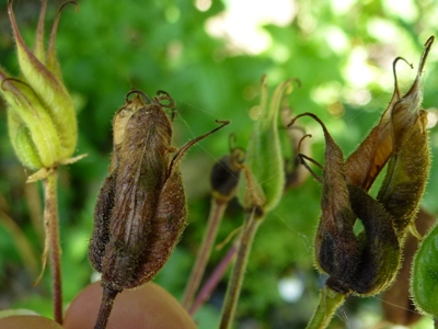
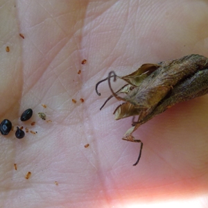
| Seed setting time, and most pods are twisted and unfilled,
possibly because of downy mildew. However, some hybrid
aquilegias find it hard to set seeds, and this could be one of
those plants. A few seeds are set in some pods, plus debris from aborted ova. Do not keep seeds from infected plants, we don't yet know if seeds could be either contaminated with or contain spores. See below, last row, center column. |
 |
 |
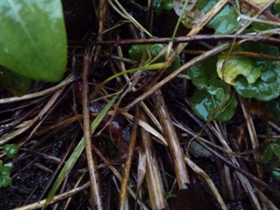
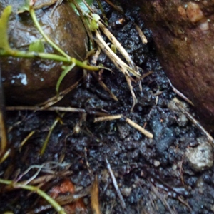
| It is now December. Winter. What's happening now? I investigated the naturally fallen stems and cleared them away to find bare soil. It looks as if this plant, too, has died unless it's completely under the soil surface at the moment. |
 |
 |
| Seeds are unlikely to carry DM. I wonder whether any spores
could be carried on the seed surface? I, and
other research establishments, are looking at DM & seeds.
Meanwhile it is very sensible not to use seeds from affected plants. I will now recommend that gardeners check ALL aquilegia seedlings for signs of downy mildew, and, if seen, get rid of the whole pot and contents. If typical lightish yellowy, angular areas appear on small seedlings' leaves, or whiter, upright growth of systemically infected seedlings, again get rid of the whole potful. Please let me know if you suspect DM in your seedlings (either self-sown ones or in pots), especially if you can take photos. |
||
Visit also the main downy mildew information page, how to spot downy mildew on leaves & more on systemic infection.
| Back to top |
Copyright Carrie Thomas 2014. Web wizardry by Trevor Rees. All rights reserved.